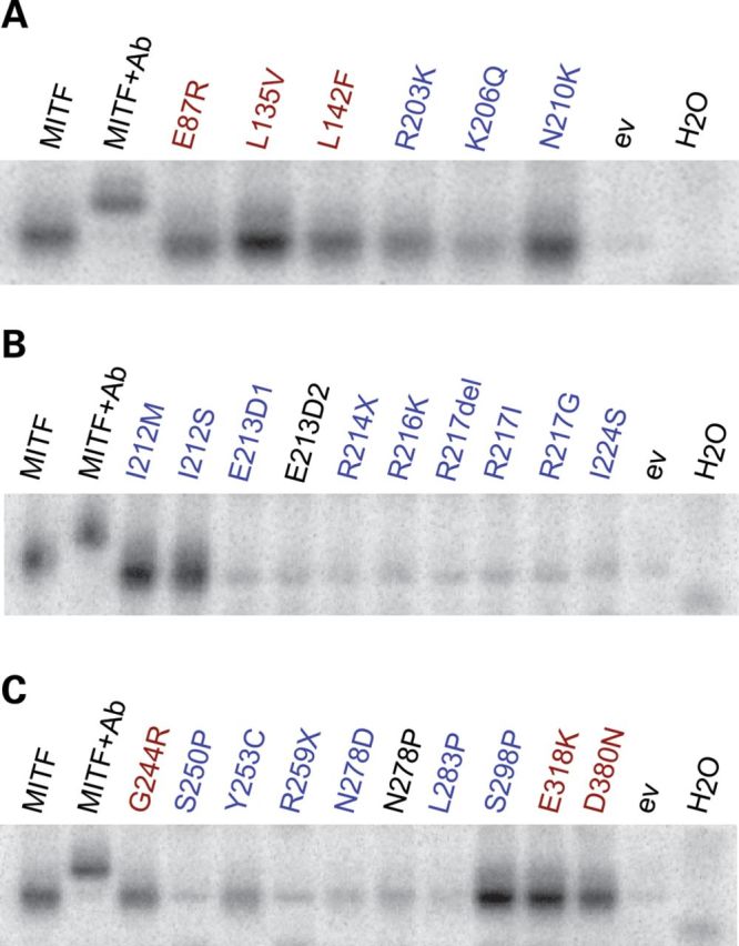
Figure 2.

MITF mutations and their ability to bind the E-box. (A–C) EMSAs showing the binding of wild-type and mutant MITF proteins to the E-box sequence (CACGTG). In each case, Lane 1 shows binding of the wild-type MITF protein, and Lane 2 shows a supershift where the C5 MITF antibody was added. Empty vector pcDNA3.1 (ev) and water (H2O) were loaded as negative controls. Melanoma mutations are indicated in red, whereas WS2A and TS mutations are indicated in blue. The synthetic E213D2 and N278P mutations are indicated in black. Representative data from three separate experiments are shown.
