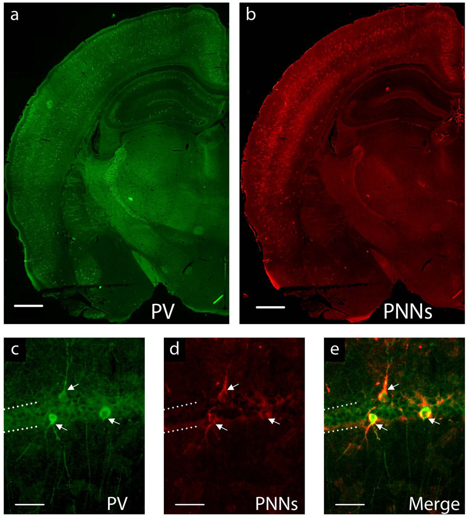
FIGURE 1.
a–b. Representative image of P65 IS whole-brain hemisphere at 4× magnification showing a. parvalbumin-positive interneurons and b. perineuronal nets. Scale bar = 1mm. c–e. Representative hippocampal CA1 interneurons at 20× magnification: c. stained with a primary antibody for parvalbumin (PV+), d. perineuronal nets (PNNs) stained with biotin-conjugated lectin from Wisteria floribunda e. Merge of images c. and d. showing PV+ interenurons surrounded by PNNs. Dotted lines indicate the CA1 hippocampal pyramidal cell layer. Arrows denote PV+ GABAergic interneurons surrounded by PNNs. Scale bar = 50µm.

