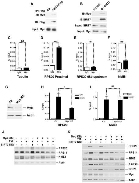
Figure 4. Myc recruits SIRT7 to repress the expression of ribosomal proteins and to suppress ER stress.
(A) Western analysis showing co-immunoprecipitation (co-IP) of Flag-tagged SIRT7 and endogenous Myc in 293T cells.
(B) Western blots showing co-IP of endogenous SIRT7 and Myc in Hep G2 cells.
(C–F) ChIP-qPCR (mean +/− S.E.M) showing Myc occupancy at the PRS20 proximal promoter but not 6kb upstream, compared to IgG negative control samples. Myc occupancy at the γ-tubulin and NME1 promoters was used as negative controls. All samples were normalized to input DNA.
(G) Western blots showing knockdown of Myc with siRNA in cells used in (H, I).
(H, I) Reduction of SIRT7 occupancy at the RPS20 but not the NME1 promoter in Myc knockdown cells determined by ChIP (mean +/− S.E.M).
(J) Western analysis showing Myc inactivation by a small molecule inhibitor abrogates increased expression of ribosomal proteins but not NME1 in stable SIRT7 KD cells used in Figure 3G.
(K) Western analysis showing Myc inactivation via siRNA abrogates ER stress and increased expression of ribosomal proteins but not NME1 in stable SIRT7 KD cells used in Figure 3G.
Error bars represent S. E. M. *: p<0.05. ***:p<0.001. ns: p>0.05.
See also Figure S3.

