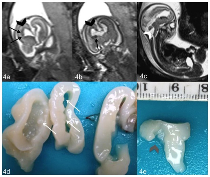
Figure 4.
Fetal brain at 20 weeks of gestation (normal male 46, XY karyotype) with bilateral subependymal heterotopia, ventriculomegaly and cerebellar abnormalities. Coronal (4a, 4b) and sagittal (4c) T2-weighted magnetic resonance images show ventriculomegaly (black arrowhead) (4a, 4b), cerebellar abnormality with a wide communication between fourth ventricle and cisterna magna (white arrowhead) (4c) and nodules of subependymal heterotopia along the walls of the frontal horns of the lateral ventricles (black arrows) (4a, 4b). Coronal and sagittal sections of the fetal brain obtained for the neuropathological examination confirmed the presence of nodules of heterotopic gray matter (white arrows) (4d) and a malrotated cerebellar vermis (gray arrowhead) (4e). (1.5 T Magnetic Resonance Imaging. Protocol: TR 1000 msec; TE 149 msec; thickness 3 mm).

