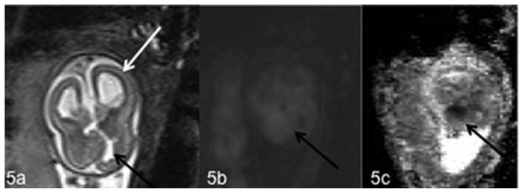
Figure 5.
Fetal brain at 20 weeks of gestation (normal male 46, XY karyotype) with bilateral subependymal heterotopia, ventriculomegaly, cerebellar asymmetry and the presence of an ischemic area. Axial T2-weighted (5a), DWI (5b) magnetic resonance images and corresponding ADC map (5c) show the presence of ventriculomegaly (white arrow) (5a) and an ischemic area on the medial surface of the frontal right lobe with mild hyperintensity on T2-weighted (5a) and DWI images (5b) and hypointensity on the corresponding ADC map (5c) (black arrows). (1.5 T Magnetic Resonance Imaging. DWI sequences Protocol: TR 8000 msec; TE 90 msec; TI 185 msec; thickness 5 mm. T2-weighted sequences Protocol: TR 1000 msec; TE 149 msec; thickness 3 mm).

