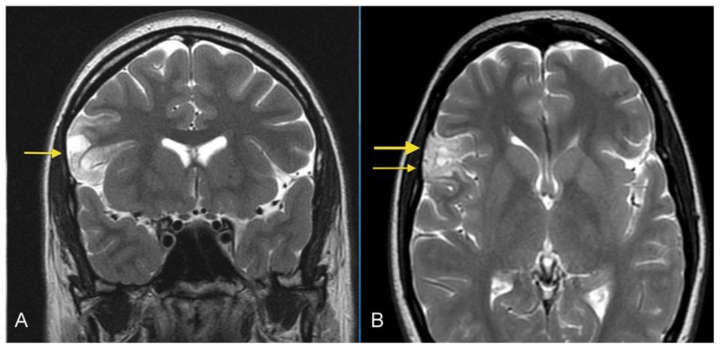
Figure 1.
26- year-old female with a dysembryoplastic neuroepithelial tumor (DNET) in the right frontal lobe. A: Coronal T2-weighted MR image shows a cortical-based superficial lesion in right frontal lobe extending to the cortical margin. There are multiple small rounded internal focal regions of high signal intensity compatible with a “soap bubble” appearance (arrow). There is no evidence of associated mass effect or vasogenic edema. (Protocol: 1.5 Tesla MRI (GE system), Repetition time/Echo time TR/TE: 3750/76.9, 3mm slice thickness, non-contrast, field of view 24 cm). B: Axial T2-weighted MR image demonstrates superficial lesion in right frontal lobe extending to cortical margin. There are internal focal regions of high signal intensity, compatible with a “soap bubble” appearance (thin arrow). There is also narrowing and remodeling of the adjacent inner table of the skull, compatible with slow growth (wide arrow). (Protocol: 1.5 Tesla MRI (GE system), TR/TE: 3200/256, 5mm slice thickness, field of view 24 cm, non-contrast).

