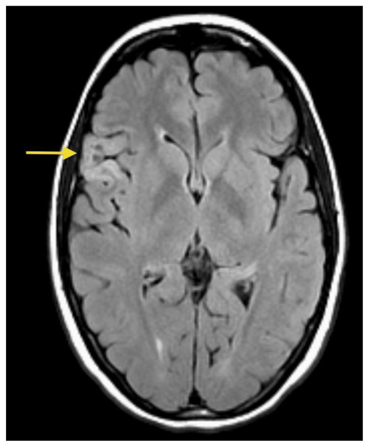
Figure 2.
26- year-old female with a dysembryoplastic neuroepithelial tumor (DNET) in the right frontal lobe. Axial FLAIR image show a slightly hyperintense cortex and slightly decreased signal in the small focal nodular areas (arrow). (Protocol: 1.5 Tesla MRI (GE system), TR/TE: 8002/123, 5mm slice thickness, field of view 24 cm, non-contrast).

