Figure 7.
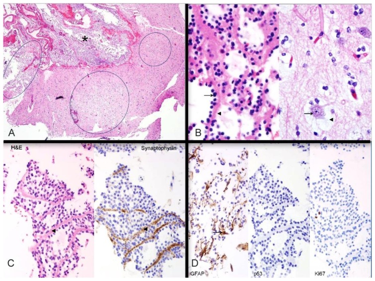
26- year-old female with a dysembryoplastic neuroepithelial tumor (DNET) in the right frontal lobe. Histochemical and immunohistochemical stains of a dysembryoplastic neuroepithelial tumor (DNET) in a 26-year-old male. A: Haematoxylin- and eosin-stained section through the tumor demonstrates discrete intracortical tumor nodules (ovals); the nodule on the left extends into subarachnoid space (asterisk). Hematoxylin and eosin (H&E), high power (40×). B: Histologic features. LEFT: Oligodendroglia-like cells (arrow) lined up along neuropil bundles (arrowhead). RIGHT: Cortical neurons (arrow) floating in myxoid pools (arrowhead). These two features comprise the “specific glioneuronal element” of dysembryoplastic neuroepithelial tumor. Both images H&E, high power (400×). C: Axonal nature of neuropil bundles. The neuropil bundles (arrowheads) in the specific glioneuronal element are immunopositive (brown) for synaptophysin, supporting their axonal nature. LEFT: Hematoxylin and eosin (H& E), high power (200×); RIGHT: synaptophysin immunostain, high power (200×). D: Additional immunohistochemical findings. LEFT: GFAP-immunopositive reactive astrocytes (arrow) are scattered in the specific glioneuronal element. CENTER: Mutant p53 protein is not expressed in the nuclei of oligodendroglia-like cells. RIGHT: Neoplastic cells show a low proliferation index with Ki-67 immunostain. Left: Hematoxylin and eosin (H&E), glial fibrillary acidic protein (GFAP) immunostain, 200×; Center: p53 protein immunostain, 200×; Right; Ki-67 immunostain, 200

