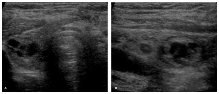
Figure 1.
Thyroid ultrasound. 71-year-old female patient with an ovarian lesion containing thyroid follicles and foci of papillary carcinoma, most probably representing a papillary carcinoma arising in a struma ovarii. Ultrasound scans in axial (1A) and sagittal (1B) planes depict multiple nodules in the right thyroid lobe, measuring up to 1.5cm in greatest axis, some of them being mixed solid and cystic and the others being predominantly cystic. None of the nodules exhibited suggestive features of malignancy [GE Voluson V730, linear transducer, 37 Hz].

