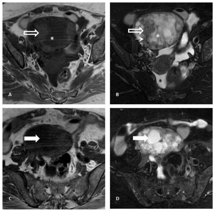
Figure 3.
Pelvic Magnetic Resonance Imaging. 71-year-old female patient with an ovarian lesion containing thyroid follicles and foci of papillary carcinoma, most probably representing a papillary carcinoma arising in a struma ovarii. Axial T1-weighted images (3A and 3C) and T2-weighted images with fat saturation (3B and 3D) revealed a multiloculated cystic mass presenting locules with variable signal intensity (“stained glass appearance”) and thick septations. Some locules showed low signal intensity on T1-weighted images and high signal intensity on T2-weighted images (white arrow; 3C and 3D), while other locules exhibited low signal intensity on both T1- and T2-weighted images (white asterisk; 3A and 3B), the latter most probably due to the presence of colloid content. Other areas with intermediate signal on T1- and T2-weighted images (white-bordered white arrow; 3A and 3B) presented linear signal voids (thin arrow; 3B) within it and were compatible with solid areas.. No fatty component was detected. A small amount of ascites was identified in the pouch of Douglas / cul-de-sac (black asterisk; 3B) [GE Signa HDe 1.5T: T1-weighted images (TR=420; TE=12,8); T2-weighted images (TR=2380; TE=105); Proton-density weighted image (TR= 3560; TE= 38,4)].

