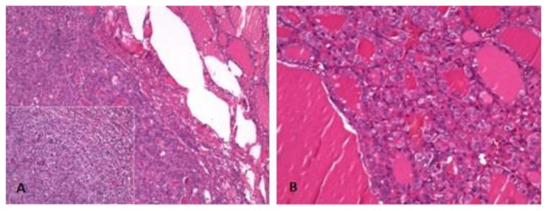
Figure 6.
Microscopic examination of hematoxylin and eosin (h&e) stained sections. 71-year-old female patient with an ovarian lesion containing thyroid follicles and foci of papillary carcinoma, most probably representing a papillary carcinoma arising in a struma ovarii. Low and high power views of left thyroid lobe depict an uncapsulated nodule with oncocytic features and empty and grooved nuclei, respectively (6A). High power view of the right thyroid lobe shows a papillary carcinoma with follicular pattern (6B).

