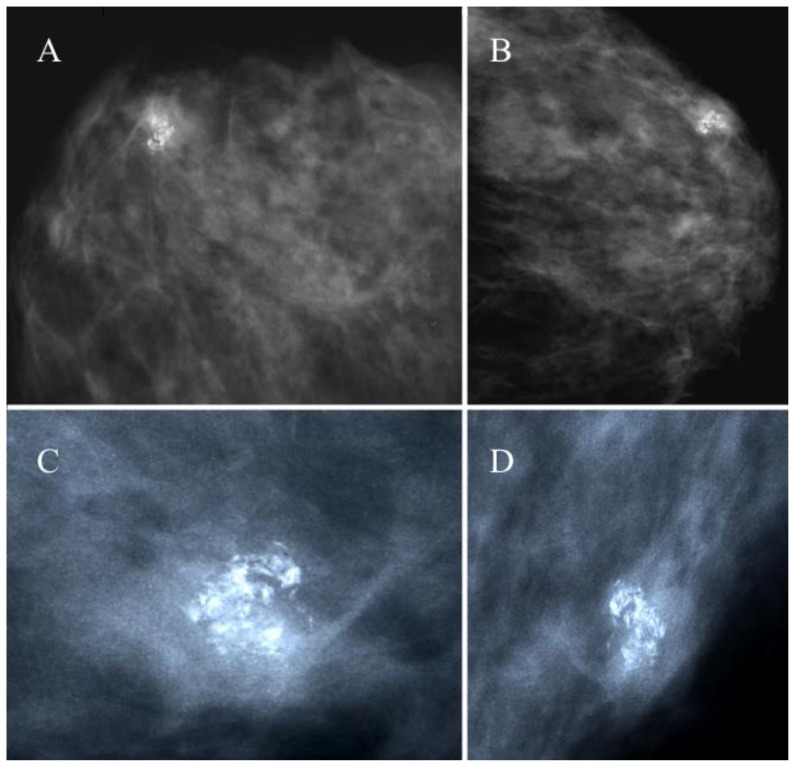
Figure 1.
43 year old female with a hard, firm nodule at the confluence of the external quadrants of the left breast diagnosed as a breast pilomatrixoma after percutaneous and excisional biopsy.
FINDINGS: nodular opacity in the external quadrants of the left breast of 12×11 mm, showing a cluster of pleomorphic irregular microcalcifications (ACR BI-RADS IV–V).
TECHNIQUE: Analogic mammography (28 kV, 100 mAs), cranio-caudal (A) and medio-lateral oblique projections (B) of the left breast. Magnification views of cranio-caudal (C) and medio-lateral oblique (D) projections showing the lesion.

