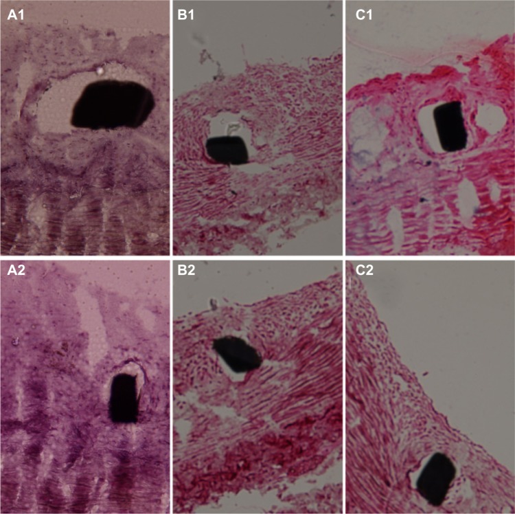
Figure 11.
Pathology of Groups A, B, and C at 2 and 4 weeks.
Notes: Two-week (A1, B1 and C1) and 4-week (A2, B2 and C2) high-power photomicrographs (H&E staining; 200 × magnification) of pathology of Groups A, B, and C. The inflammation response was significant lower in Groups A and B than in Group C.

