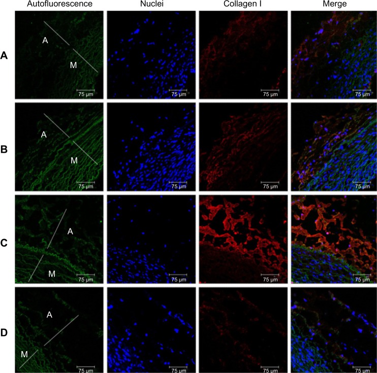
Figure 13.
Immunofluorescence of collagen I close to stented arteries.
Notes: Collagen type I immunostaining (orange) of acetylsalicylic acid-loaded (Groups A and B) and non-acetylsalicylic acid-loaded (Group C) nanofibrous membrane stent. (Group D) normal vessels. Autofluorescence on tunica media (green), and DAPI-stained nuclei are also shown. Less collagen type I-positive labeling was observed close to the acetylsalicylic acid-loaded stent vessels. scale bar: 75 μm.
Abbreviations: A, tunica adventitia; M, tunica media; D, control group.

