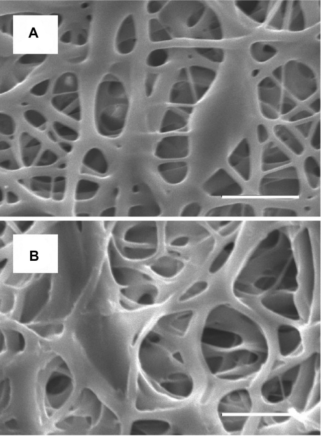
Figure 2.

Morphology of nanofibrous membrane elucidated by scanning electron microscopy.
Notes: Images of acetylsalicylic acid-loaded nanofibers within the diameter range 50–8,720 nm, (A) before expansion (the pore size is around 5 μm), and (B) after expansion by a balloon (the pore size is around 10 μm). Scale bar: 10 μm.
