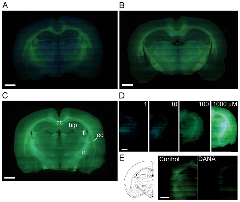
Figure 4. Visualizing of sialidase activities in rat brain slices with BTP-Neu5Ac.
A–C, Sialidase activity was imaged in acute coronal slices of adult rat brains with BTP2-Neu5Ac (A), BTP3-Neu5Ac (B) and BTP4-Neu5Ac (C) at pH 7.3. Abbreviations: cc, corpus callosum; ec, external capsule; fi, fimbria; hip, hippocampus; ic, internal capsule. D, Brain slices were stained with various concentrations (1–1000 µM) of BTP4-Neu5Ac. E, Sialidase activity was imaged with 10 µM BTP4-Neu5Ac or 10 µM BTP4-Neu5Ac containing 1 mM DANA, a sialidase inhibitor. Emission filters that transmit above 420 and 510 nm were used in A–D and E, respectively. Scale bars in each panel represent 2 mm.

