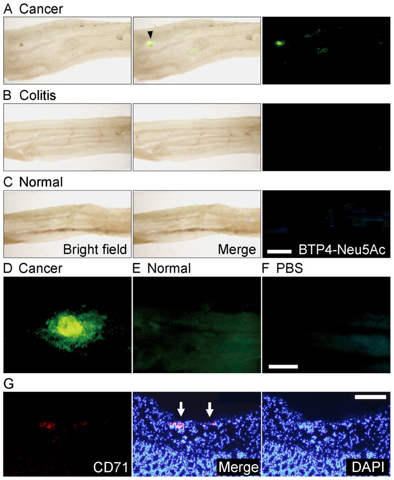
Figure 6. Detection of orthotopic colon cancer with BTP4-Neu5Ac in mouse colon tissue.
A, One week after orthotopic colon implantation of Colon26 NL-17 cells, living colon tissues were stained with BTP4-Neu5Ac. Arrowhead indicates the cancer region. B and C, Inflammatory (B) or normal (C) colons were also stained with BTP4-Neu5Ac. Left, middle and right panels in panel A–C show bright field, merged and fluorescent views, respectively. D and E, Enlarged image of cancer (D) and normal (E) region stained with BTP4-Neu5Ac. F, Background fluorescence level of panels D and E is shown. G, Immunohistochemical staining (red fluorescence) by using rabbit anti-CD71 antibody and PE-conjugated goat anti-rabbit IgG antibody and nuclear staining with DAPI (blue fluorescence) were performed to detect colon cancer in cross sections of the mouse colon tissues that were used for sialidase activity imaging in panels A and D. Arrows indicate the regions showing intense fluorescence of BTP in panel A and D. Scale bar in panel C represents 2.5 mm and is common in panels A and B. Scale bar in panel F represents 0.5 mm and is common in panels D and E. Scale bar in panel G represents 0.2 mm.

