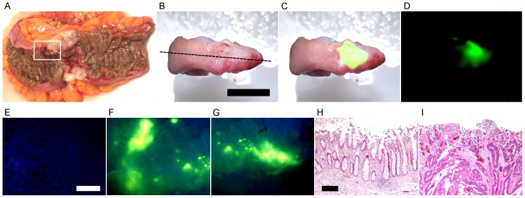
Figure 7. Detection of human colon cancer with BTP4-Neu5Ac.
A, A human colon cancer specimen (region enclosed with a white line) was obtained from surgical cancer tissue (UICC T classification: T3). B–D, The colon tissue was stained with BTP4-Neu5Ac. B, C and D show photographic, merged and fluorescent images, respectively. E–G, Enlarged fluorescence images of normal (E) and cancer (F and G) regions were acquired with a fluorescent microscope. H and I, A longitudinal slice of colon tissue was prepared at the dotted line in panel B. Non-fluorescence and fluorescence regions in panel C were stained with hematoxylin-eosin and are shown in panels H and I, respectively. Scale bars in panel B, E and H represent 10 mm (common in panels B–D), 500 µm (common in panels E–G) and 250 µm (common in panels H and I), respectively.

