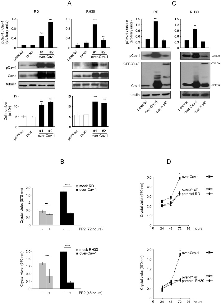
Figure 2. Effects of Cav-1 overexpression on cell proliferation.
RD and RH30 cells were stably transfected with either Cav-1 construct (over-Cav-1) or empty vector (mock). A) Parental cells and selected clones were seeded in 60-mm dishes (at a density of 15×104). After 48 hours, cells were harvested and protein homogenates western blotted for pCav-1, Cav-1 and tubulin. Protein bands were quantified by densitometry after normalization with respect to tubulin (n = 3). **, P<0.001; ***, P<0.0001. At the same time points, cell proliferation was evaluated with Burker chamber, as shown in the bottom graphs. Bar graphs represent means ± SD of total cell numbers. (n = 3) ***, P<0.0001. B) Mock and over-Cav-1 cells were seeded in 24-well plates (at a density of 15×103). After 24 hours, cells were either treated with dimethylsulfoxide (DMSO, vehicle) or 10 µM PP2 (replenished every 24 or 16 hours for RD and RH30 cells, respectively) for the indicated time points. Cell proliferation was then evaluated by Crystal violet assay. Histograms represent means ± SD of absorbance (n = 4). ***, P<0.0001; ****, P<2e-16. C) RD and RH30 cells were stably transfected with constructs for either Cav-1 (22 kDa) or non-phosphorylatable GFP-Y14F (55 kDa). Parental cells and selected clones were seeded in 60-mm dishes (at a density of 12×104). After 24 hours, cells were harvested and protein homogenates western blotted for pCav-1, Cav-1 and tubulin. Protein bands were quantified by densitometry after normalization with respect to tubulin (n = 3). **, P<0.001; ***, P<0.0001. D) Under the same conditions seen above, cell proliferation was evaluated by Crystal violet assay at the indicated time points. Histograms represent means ± SD of absorbance (n = 4). *, P<0.05.

