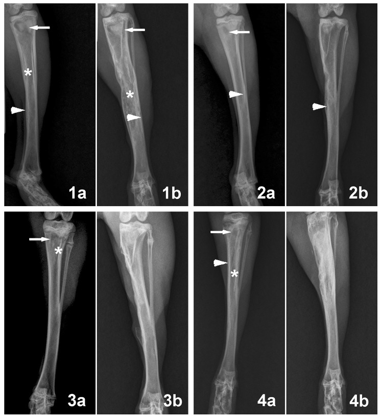
Figure 7. Radiographics analysis of animals before surgery and at 2 months post-surgery.
Representative preoperative radiographs showing tibial osteomyelitis in the four different groups (1a, 2a, 3a, 4a), characterized by bone destruction (arrows), periosteal new bone formation (arrowhead) and sequestral bone formation (asterisk). (1b) Postoperative radiographs of Group 1 showed deterioration of osteomyelitis coupled with periosteal new bone formation (arrowhead), destruction of bone (arrows) and sequestral bone formation (asterisk). (2b) Postoperative radiographs of Group 2 showed that the osteomyelitis was partly controlled. (3b, 4b) Postoperative radiographs of Group 3 and Group 4 showed that the osteomyelitis was healed.

