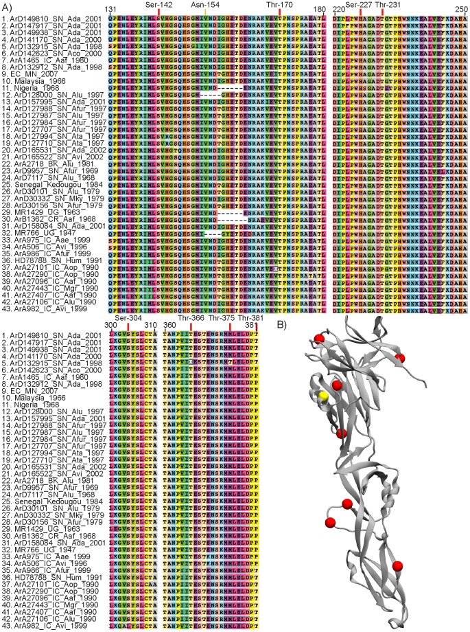
Figure 3. Mapping of predicted glycosylation sites on envelope protein of ZIKV.
A) Alignment of E protein showing predicted glycosylation sites. Red arrows point to O-linked glycosylation sites (Ser or Thr residues) and the yellow arrow points to the N-linked glycosylation site (Asn-X-Thr motif). B) Tridimensional structure of E protein. Red beads indicate O-linked glycosylation sites and the yellow bead indicates the unique N-linked glycosylation site.

