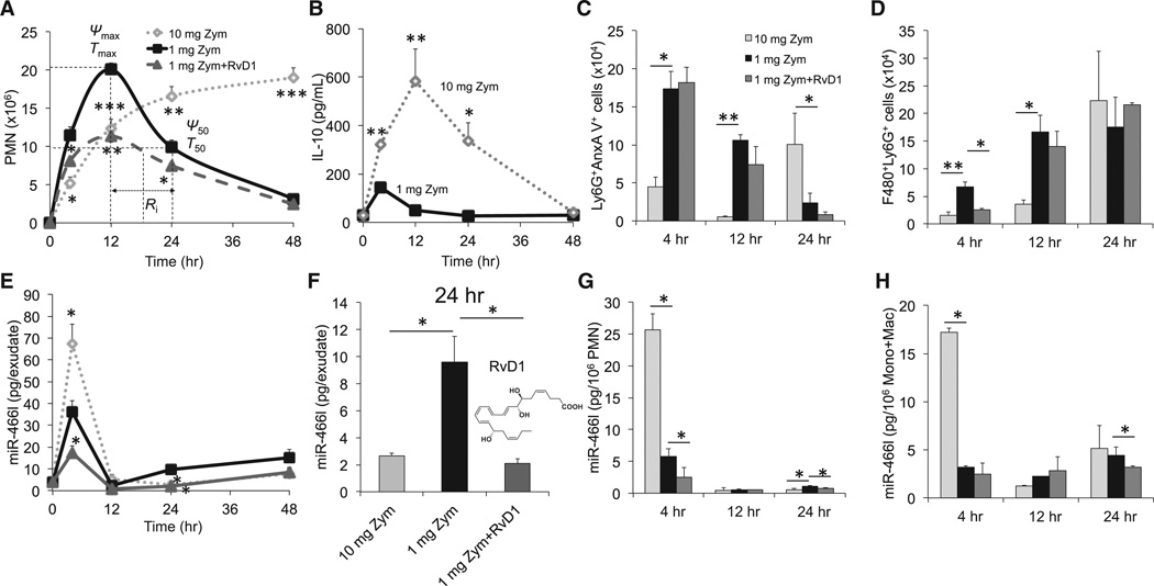
Figure 1. Temporal and Differential Regulation of Exudate Leukocyte miR-466l during Self-limited versus Delayed Inflammation Resolution.
Zym was injected (i.p.) for acute peritonitis into male FVB mice divided into three groups: high-dose (10 mg/mouse) challenged, low-dose (1 mg/mouse), and RvD1-treated.
(A) Lavages were collected at indicated intervals, and PMNs were enumerated. See Experimental Procedures and Table S1 for the calculation of resolution indices.
(B) Exudates from 1 and 10 mg of Zym-initiated murine peritonitis (0–48 hr) were collected for IL-10.
(C) Flow cytometry for apoptotic PMNs (Ly6G+AnxAV+).
(D) Flow cytometry for efferocytosis (F4/80+Ly6G+).
(E) Total RNAs were isolated at the indicated intervals, and expression of miR-466l was assessed by quantitative PCR (qPCR).
(F) miR-466l expression in each group at 24 hr.
(G and H) Quantitative expression of miR-466l in FACS-sorted PMNs (G), monocytes, and macrophages (H) from murine exudates. Results are expressed as mean ± SEM from at least four independent experiments for each point in each panel. *p < 0.05, **p < 0.01, and ***p < 0.001 versus low-dose group.
See also Figure S1 and Table S1.

