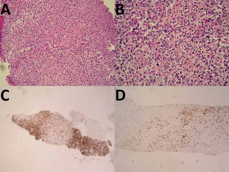
Figure 2.
Histopathology of mass at caudate lobe show necrotic tissue with atypical plasma cell-like infiltration (A and B). Immunohistochemistry show CD3: negative, CD20: positive, (C) Ki67: 30–40% positive, CD79a: positive, CD23: negative, CyclinD1: negative, CD10: negative, Bcl-6: negative, MUM1: positive, BCL2: positive, EBV (LMP): positive (D), HHV8: negative and EBER: positive. B-cell lymphoma-6; BCL-2, B-cell lymphoma-2; EBV, Epstien-Barr virus; HHV8, human herpes virus 8; MUM1, multiple myeloma oncogene 1; EBER, EBV-encoded RNA.

