Figure 3.
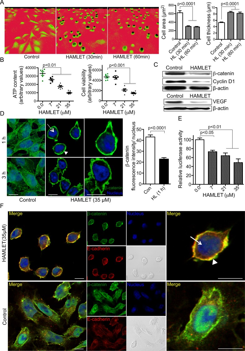
Effects of human α-lactalbumin made lethal to tumour cells (HAMLET) on Wnt/β-catenin pathway in vitro. APC mutated, DLD1 human colon cancer cells were exposed to HAMLET. (A) Time-dependent morphological changes, detected by phase contrast holographic microscopy. Significant reduction in surface area (p<0.0001) and increase in thickness (p<0.0001) are quantified (one-way analysis of variance (ANOVA) test, n=3). (B) Dose-dependent loss of viability after HAMLET treatment (3 h), quantified by ATP measurements (p<0.01) and Presto blue (p<0.001) (one-way ANOVA test, n=3). (C) Decrease in β-catenin, cyclin D1 and VEGF protein levels after HAMLET treatment (3 h, 35 µM). Western blot, with β-actin as loading control. (D) DLD1 cell monolayers were treated with HAMLET (35 µM, 1 and 3 h), fixed and immunostained for β-catenin (green). Nuclei were counterstained with DRAQ5 (blue). HAMLET significantly reduced nuclear β-catenin staining in treated cells (p<0.0001) (t test, n=3). The uniform cytoplasmic staining was replaced by strong membrane staining (scale bars, 10 μm). (E) TCF/LEF (T-cell factor/lymphoid enhancer binding factor) reporter activity quantified by TOP-flash dual luciferase assay. A dose-dependent reduction in luciferase activity was detected after 3 h of HAMLET treatment (p<0.01 and p<0.05) (one-way analysis of variance test, n=3). (F) HAMLET enhances colocalisation of β-catenin and E-cadherin on the cell membrane. Cells exposed to HAMLET (35 μM, 3 h) were double stained for β-catenin (green), E-cadherin (red) and DRAQ5 (blue) as a nuclear staining agent. Both β-catenin and E-cadherin were intensely colocalised at the cell membrane with increase in β-catenin staining (scale bars, 10 μm). In all panels, error bars represent±SEM.

