
Figure 3.
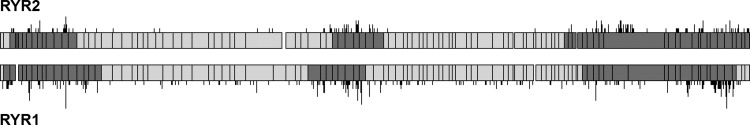
Disease-causing variation in human ryanodine receptors, RYR1 and RYR2. An alignment of RYR1 and RYR2 reveals the structural similarity of the proteins and homologous clustering of pathogenic variation in these related proteins. The protein is represented in light grey, with reported ‘mutation hotspots’ marked in dark grey, and exon boundaries highlighted. The locations of missense variants previously reported to be pathogenic are shown with black lines above and below the protein graphic—longer lines indicate more than one pathogenic DNA variant affecting the same protein residue.