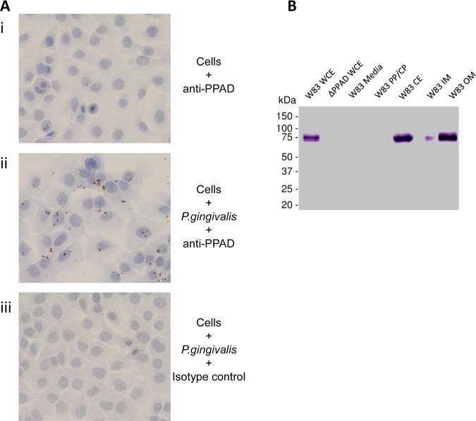
Figure 2.
PPAD associates with the bacterial cell and is expressed on the outer membrane (OM). (A) An immortalised oral keratinocyte cell line (OKF6) was stained with anti-PPAD antibody (i). Cells were infected with Porphyromonas gingivalis and stained with anti-PPAD antibody (ii) or isotype control (iii). Cell nuclei were counterstained blue. Magnification: ×400. (B) PPAD subcellular localisation in P gingivalis. Bacteria culture from an early stationary phase was fractionated into whole cell extract (WCE), particle-free growth media (Media), soluble cell proteins derived from periplasm and cytoplasm (PP/CP), cell envelope containing inner and outer membranes (CE), inner membrane (IM) and outer membrane (OM). Fractions were subjected to Western blot analysis using anti-PPAD antibody.

