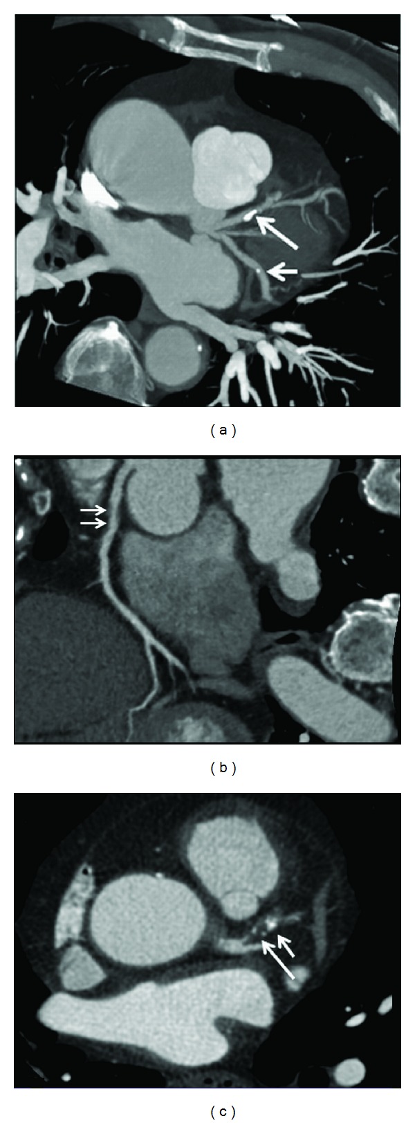
Figure 3.

Maximum-intensity projection shows that calcified plaques are present at the proximal segment of left anterior descending (long arrow) and midsegment of left circumflex (short arrow) (a). Curved planar reformation shows a noncalcified plaque at the proximal segment of right coronary artery ((b) arrows). 2D axial image demonstrates a mixed plaque at the proximal segment of left anterior descending (c), with short arrow referring to the calcified component and long arrow to the noncalcified component within the plaque.
