Figure 8.

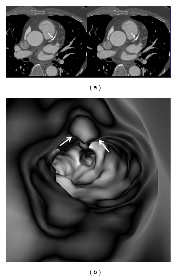
A heavily calcified plaque is present at the proximal segment of left anterior descending ((a) arrows). VIE shows irregular intraluminal appearance ((b) arrows).

A heavily calcified plaque is present at the proximal segment of left anterior descending ((a) arrows). VIE shows irregular intraluminal appearance ((b) arrows).