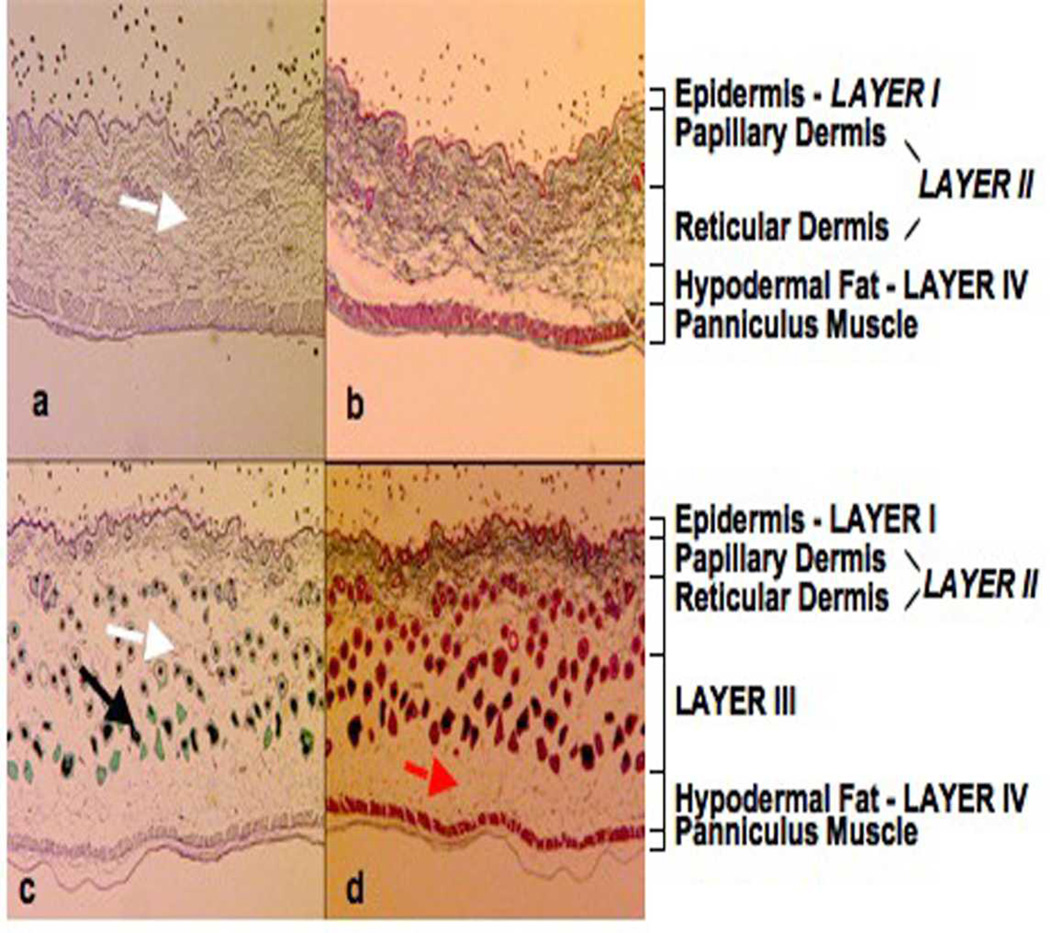
Figure 1.
Luna (a and c) Masson's trichrome (b and d) histological staining of mouse skin from the dorsum of wildtype (+/+) and homozygous oim/oim mice. The Masson’s trichrome stain demonstrated a much reduced region of collagen-rich tissue in the oim/oim compared to the +/+ animals and an increase in dermal adipose tissue (red arrow). The Luna stain showed a loss of elastin fibers (white arrow) in the lower reticular dermal layer (lower part of layer II and layer III) of the oim/oim compared to the +/+ skin samples. In the oim/oim mouse many hair follicles were present that were in the anagen stage of hair growth, with large, heavily pigmented, melanin-rich, hair bulbs. The hair follicles (black arrow) had thick root sheaths, while sebaceous glands were small and inconspicuous. Epidermis – Layer I, Papillary Dermis and Reticular Dermis – Layer II and Hypodermal Fat – Layer IV.

