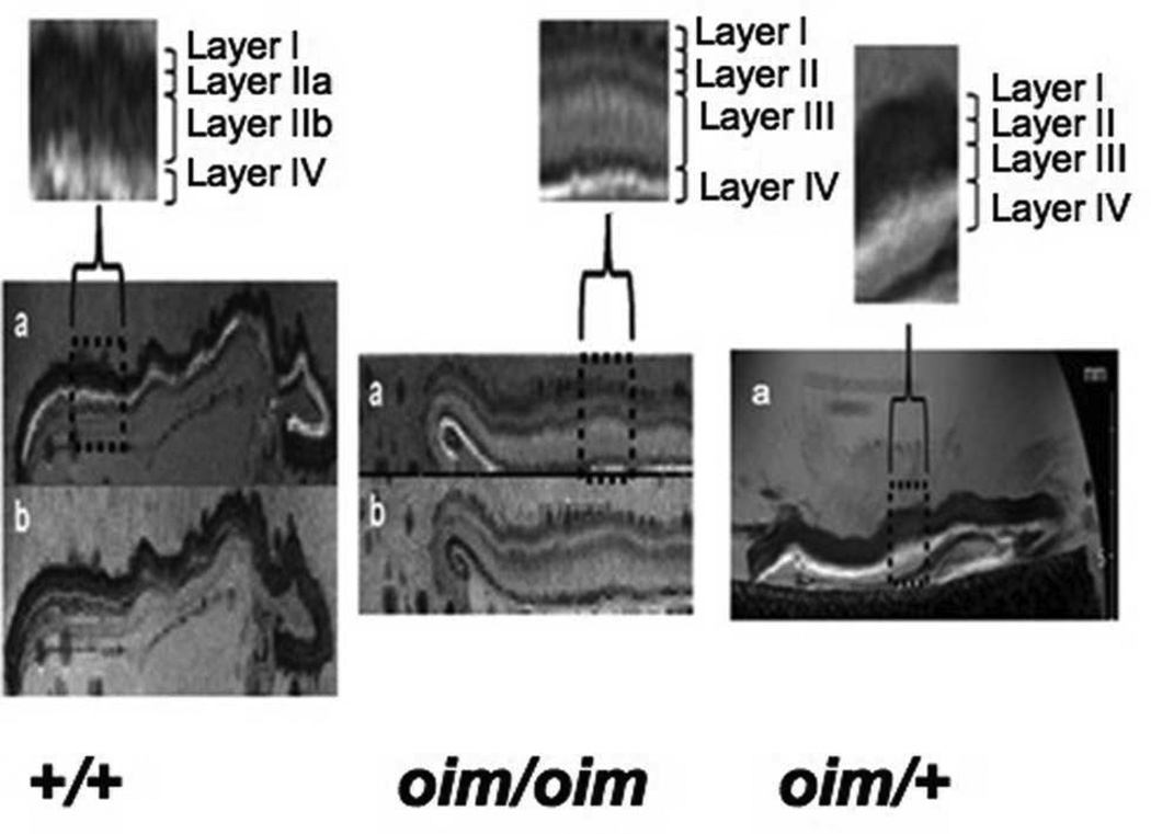
Figure 2.
T2-weighted images (a) showed a clear demarcation of individual skin layers in the wildtype control (+/+) and homozygous (oim/oim) mice skin samples. The fat suppression techniques (TE = 12 ms, TR = 15 s, NEX= 2) caused the hypodermal fat layer to be darkened (b) thus permitting the identification of any fatty layers in the MR image. Differentiation of the reticular dermal layer into an upper (layer II) and a lower layer (layer III) was seen in all the oim/oim skin samples, and in one out of the four heterozygous (oim/+) skin samples examined (pictured). Layers lla and llb correspond to the papillary and reticular dermis, respectively.

