Fig. 6.
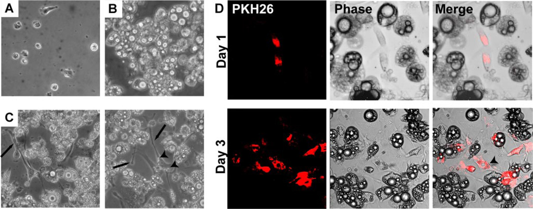
Direct coculture of macrophages with adipocytes alters macrophage morphology. A: phase contrast pictures of thioglycollate PMs in isolated culture. B: phase contrast pictures of differentiated 3T3-L1 adipocytes in isolated culture. C: direct coculture of PMs and adipocytes after 3 days demonstrates that macrophages take on an elongated phenotype with long cellular processes (arrows). Two representative views are shown. Increased accumulation of intracellular lipid (arrowhead) is also seen in macrophages. D: PMs were labeled with PKH26 (red) prior to plating on differentiated 3T3-L1 adipocytes. After 1 day of coculture with adipocytes (top), MPs appear small and oblong. After 3 days of coculture (bottom), PKH26+ macrophages appear more elongated and accumulate intracellular lipid (arrowhead).

