Fig. 3.
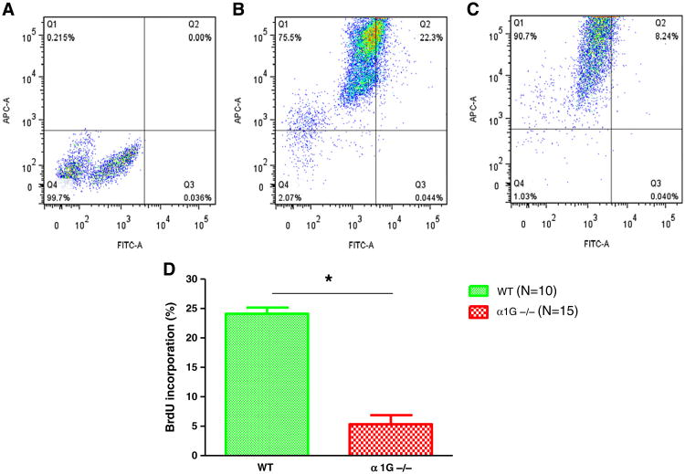
BrdU incorporation into DNA of NMVMs during the first week after birth determined with flow cytometry. NMVMs were isolated from BrdU injected mouse hearts 7 days after birth. NRVMs were stained for cardiac actin-APC and BrdU-FITC to identify myocytes within newly formed DNA. (A) Unstained control NRVMs were used to set thresholds for identifying NRVMs with BrdU+ DNA. This approach was confirmed versus studies with animals that had not been BrdU injected, but myocytes were stained for BrdU (see Supplemental Fig. 1). (B) In wild type NMVMs 22.3% were BrdU+. (C) In α1G−/− NMVMs 8.24% were BrdU+. (D) Average data from 10 WT and 15 α1G−/− NRVM preparations. There were significantly more BrdU+ myocytes in WT versus α1G−/− myocytes. *p < 0.05 (N: # of hearts).

