Fig. 8.
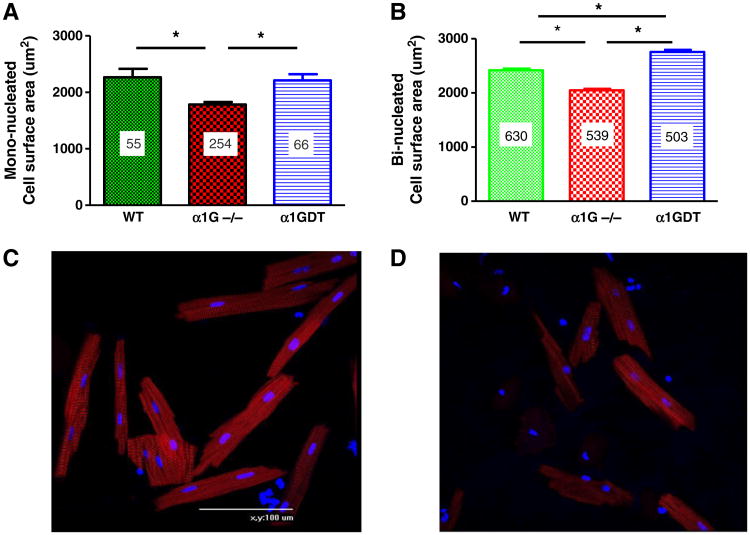
Cell surface area was measured in myocytes isolated from wild type (N = 3), α1GDT (N = 3) and α1G−/− (N = 3) 2 months old mice. (A) Cell surface area of mono-nucleated myocytes in wild type (n = 55) and in α1GDT (n = 66) was larger than in α1G−/− (n = 254). (B) Cell surface area of bi-nucleated myocytes from wild type (n = 630) and α1GDT (n = 503) hearts were larger than in α1G−/− (n = 539). (C) Representative wild type and (D) α1G−/− myocytes from 2 month old hearts are shown. Cardiac α-actin is in red and DAPI is in blue. *p < 0.0001, n: # of myocytes, N: # of hearts.

