
Figure 1.
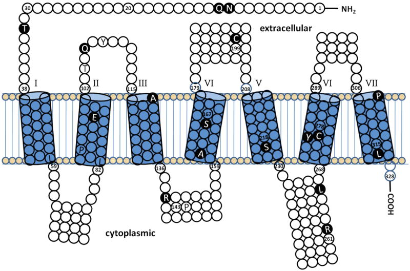
Shown is a topology diagram of the amino acid sequence of the human GnRHR protein that was generated using the HMMTOP transmembrane topology prediction server [53, 54]. The predicted seven transmembrane domains are indicated as cylinders, numbered 1-7. Also shown are the predicted extracellular loops above and the intracellular loops below the transmembrane domains. All missense mutations confirmed by in vitro analysis and one nonsense mutation (L314X) are depicted in white amino acid abbreviations on black circles. Missense mutations not studied in vitro are depicted in black letters on white or shaded circles. Residue numbers are given at the boundary of transmembrane domains and at other locations near mutated residues.