
Fig. 8.
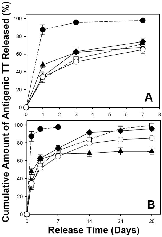
Effect of encapsulation methods (traditional w/o/w emulsion-solvent evaporation vs. active self-healing encapsulation), type of plasticizer (slightly water-soluble diethyl phthalate (DEP) vs. insoluble tributyl acetylcitrate (TBAC)), and mixed porosigens (3 wt% trehalose vs. 1.5 wt% trehalose + 1.5 wt% MgCO3) on initial burst (A) and long-term (B) in vitro antigenic TT release kinetics from PLGA microspheres. The TT release kinetics were from unencapsulated Al(OH)3 (●), TT/PLGA microspheres prepared from w/o/w emulsion solvent evaporation (▲), and TT/adjuvant/plasticizer/PLGA microspheres after self-healing microencapsulation (□: 3.2 wt% Al(OH)3/3.5 wt% trehalose/5 wt% DEP/PLGA microspheres; ◆: 3.2 wt% Al(OH)3/3 wt% trehalose/5 wt% TBAC/PLGA microspheres; ○: 3.2 wt% Al(OH)3/1.5 wt% trehalose/1.5 wt% MgCO3/5 wt% TBAC/PLGA microspheres). The release curves with dashed line are reproduced from [32] for comparison. In vitro release was evaluated in PBST + 0.2% BSA at 37 °C, and symbols represent mean ± SEM, n = 3.