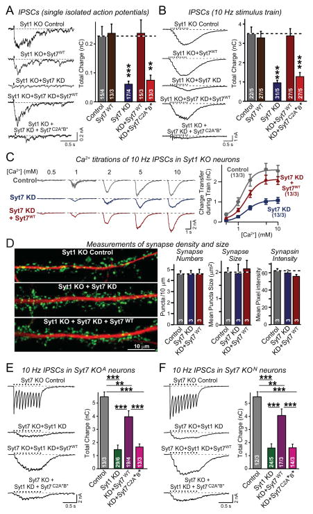
Figure 3. Suppression of asynchronous release by Syt7 KD or KO is rescued by WT but not mutant Syt7.
A & B, Syt7 KD impairs asynchronous release (monitored as IPSCs) induced by isolated action potentials (A) and action-potential trains (B) in Syt1 KO neurons. This impairment is rescued by WT Syt7 (Syt7WT) but not by mutant Syt7 containing substitutions in the C2A- and C2B-domain Ca2+-binding sites (Syt7C2A*B*). Cultured hippocampal neurons were infected at DIV4 with control lentivirus, lentivirus expressing WT Syt7 without shRNAs, or lentivirus expressing the Syt7 KD without or with shRNA-resistant WT or mutant Syt7 rescue cDNAs. At DIV14-16, IPSCs were recorded after stimulation by isolated action potentials or by trains of action potentials (10 Hz, 1 s).
C, Ca2+-titrations of asynchronous release in Syt1 KO neurons infected with control lentivirus or lentiviruses expressing the Syt7 KD without or with WT Syt7 rescue. IPSCs elicited with a 10 Hz 1 sec stimulus train were monitored at the indicated concentrations of extracellular Ca2+ (left, representative traces; right, plot of the synaptic charge transfer as a function of the extracellular Ca2+-concentration).
D, Syt7 KD does not alter synapse density or size. Control Syt1 KO neurons or Syt1/Syt7 double-deficient neurons without and with expression of WT Syt7 (obtained as in A) were analyzed by double immunofluorescence labeling for synapsin (green) and MAP2 (red; left = representative images; right = quantifications of synapse number, size, and synapsin staining intensity).
E & F, Impaired asynchronous release in Syt7 KO/Syt1 KD neurons is rescued by WT but not mutant Syt7. Hippocampal neurons cultured from two independent Syt7 KO mouse lines (E, Syt7 KOA from Maximov et al., [2008]; F, Syt7 KON from Chakrabarti et al. [2003]) were infected with control lentivirus or Syt1 KD lentivirus without or with superinfection with a second lentivirus expressing WT or mutant Syt7. IPSCs were elicited by 10 Hz, 1 sec stimulus trains (left, representative traces; right, summary graphs of the total synaptic charge transfer). Syt7 KO neurons exhibit normal synchronous release (control) that is blocked by the Syt1 KD, but in Syt1/Syt7 double-deficient neurons, asynchronous release is also largely absent but can be selectively restored by expression of WT Syt7.
All data are means ± SEM; numbers in bars indicate number of neurons/independent cultures or number of independent cultures analyzed. Statistical significance was assessed by one-way ANOVA (**, p<0.01; ***, p<0.001) comparing test conditions to control (A–D) or to the non-Syt1 KD and the WT Syt7 rescue condition (E and F). For more data, see Figs. S3 and S4.

