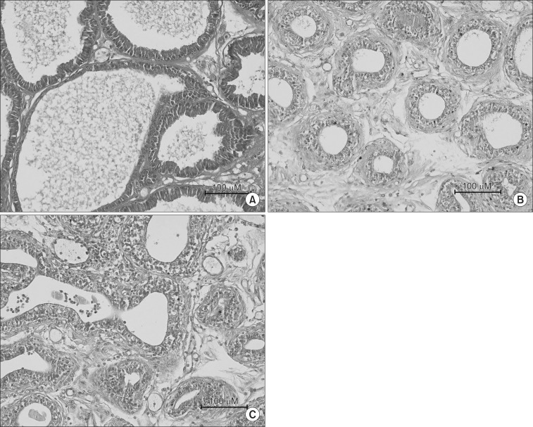
Fig. 2.
H&E stain of the prostate in each experimental groups. (A) It shows normal prostate gland (Group I). (B) All acini of the prostate gland were diffusely atrophic. Each atrophic acini formed a relatively certain round shape and were separated by thick fibrohyaline collar and stromal fibrosis (Group II). (C) All acini of the prostate gland were diffusely atrophic. The variable sized and shaped acini closely packed together and lined by atrophic epithelium. Also fibrohyaline collar and stromal fibrosis, separated each acini, were decreased (Group III).

