Figure 3.
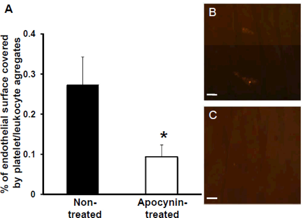
Assessment of platelet adhesion on the aortic endothelial surface after 7 days of saline or apocynin treatment. A, Percentage of the endothelial surface of the ascending aorta covered with platelet/leukocyte aggregates (n=5 in each group), *P<0.05 vs nontreated animals. Examples of en face fluorescence microscopy demonstrating 2 platelet/leukocyte aggregates on normal appearing endothelial surface in a nontreated animal (B) and absence of platelet/leukocyte aggregates in an apocynin-treated animal (C). Scale bar, 25 µm.

