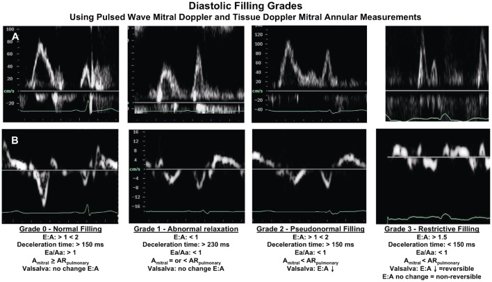
Figure 2. The stages of diastolic dysfunction recognized by changes in left ventricle filling dynamics.
(A) Pulsed-wave Doppler of mitral inflow and (B) Pulsed-wave tissue Doppler of mitral annulus in progressive stages. Diastolic filling grades: classification of diastolic filling and representative mitral inflow pulsed wave Doppler and mitral annulus pulsed wave tissue Doppler signals; E/A: ratio of passive early to active late mitral filling velocities; Deceleration time: time from peak to baseline of the mitral E velocity; Ea/Aa: ratio of the early mitral annular velocity to the late annular velocity; Amitral: duration of filling of the late filling velocity of the mitral inflow; ARpulmonary: duration of atrial reversal component of the pulmonary venous inflow. Statement: this figure was adapted from Whalley GA, Wasywich CA, Walsh HJ, Doughty RN. The role of echocardiography in the contemporary management of heart failure. Expert Rev Cardiovasc Ther 2005; 3(1): 51–70, which was authorized by the publisher.

