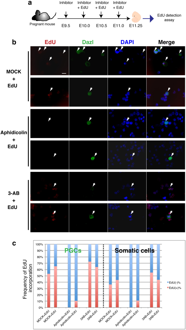
Figure 2. Small molecule inhibitor assay in the fetus during embryonic development.
(a) Experimental scheme of the inhibitor assay (drawings by Kawasaki et al.). (b) EdU detection in PGCs and somatic cells in the genital ridges from an inhibitor-treated fetus. The incorporated EdU (red) was detected in PGCs and somatic cells. DAPI was used for nuclear staining (blue). For the identification of PGCs, gonad cells were stained with Dazl (green; arrows indicate PGCs). Scale bar = 20 μm. EdU was incorporated in the 3-AB-treated or MOCK fetal genomic DNA, but not in the aphidicolin-treated fetal genomic DNA. (c) Percentage of EdU (−) and EdU (+) cells of each group of PGCs and somatic cells was presented.

