Fig. 3.
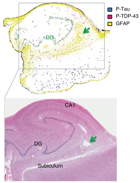
Histopathology of HS-Aging: Composite low-power figure depicts the distribution of pathology localized with multiple pathological immunomarkers. Sections were analyzed from the brain of a man who died cognitively impaired at age 92 years; autopsy showed HS-Aging and Braak stage II pathologies. An Aperio ScanScope XT with Genie™ image recognition software was used to highlight the positive immunoreactivity. The top portion shows the composite results of three nearly consecutive sections stained for phospho-tau (blue), phospho-TDP-43 (red); and GFAP (yellow). Labeled are dentate granule cell layer (DG, shown in green in top portion), CA1, and subiculum (bottom). Green arrows show same abnormally enlarged Virchow-Robin space on both top and bottom figures. This representative case shows that HS-Aging brains have a multifaceted pathological picture that includes TDP-43 pathology, astrocytosis, tauopathy, and vascular profiles that are aberrant in comparison to that which would be observed in younger control individuals. Future work is required to identify the truly specific, and clinical disease-driving, feature(s) of HS-Aging pathology

