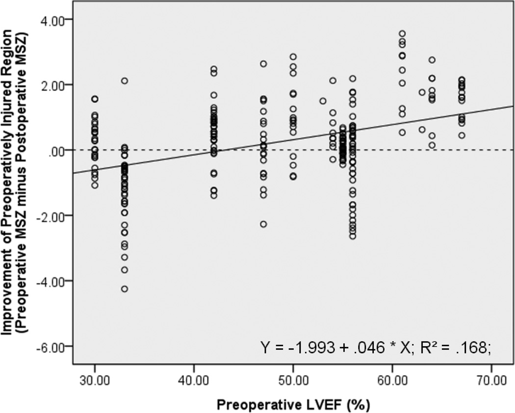
Figure 6. Regional Improvement by LVEF.
For 271 preoperatively impaired regions (from 14 AI patients), the preoperative MRI-based LVEF from the ventricle in which each region resides is significantly correlated with the improvement of that specific region following AVR (r = .410, p < .001), though not as well the global average MSZ is (p < .001). Y-Axis: ‘0’ = no improvement; positive values are improvement; negative values mean the region is faring worse after surgery.

