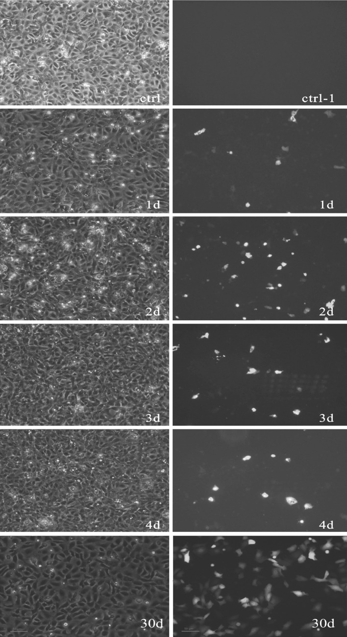
Fig. 1.
pEGFP-N1-VP6 expressed in CIK cells. Images were taken with white light (left) and fluorescent light (right). CIK cells expressing green fluorescent protein on different days (day 1, 2, 3 and 4) post transfection with pEGFP-N1-VP6 were examined by microscopy and fluorescent microscopy (control–4d) and the CIK cells transformed with pEGFP-N1-VP6 were screened by G418 in 1 month (30d)

