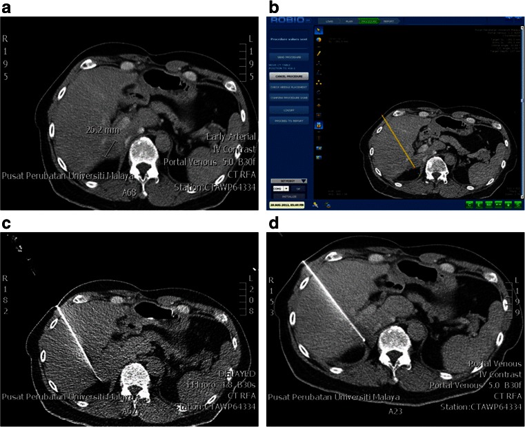
Fig. 1.
a Contrast-enhanced baseline CT image shows solitary colorectal metastases (26.2 mm diameter) in segment VI. b Reconstructed CT images (slice thickness 1 mm) were sent to the ROBIO™ EX workstation for treatment planning. The simulated needle trajectory path was shown on the treatment plan and verified by the radiologist. c A CT fluoroscopy check was carried out to verify the accuracy of the needle placement within the target volume. d Post-RFA three-phase CTs to assess the completeness of tumour ablation

