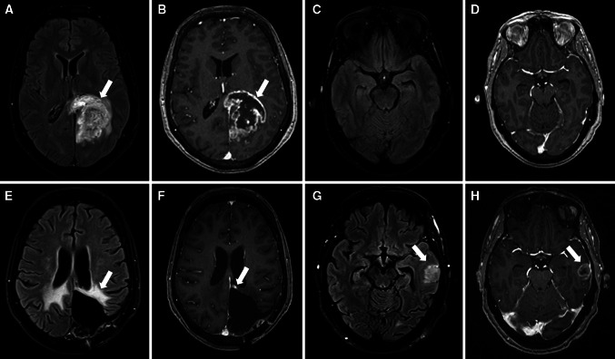
Fig. 1.
63 years-old female with GBM. MRI at diagnosis (upper row) and first recurrence (lower row). At diagnosis a left parieto-occipital mass was found, hyperintense in T2 FLAIR (arrow in a) and with irregular peripheral enhancement with central necrosis in T1Gd (arrow in b), with normal left temporal lobe (c and d). 13 months after surgery there was no evidence of recurrence at the surgical cavity borders with only treatment-related changes, with gliosis in T2 FLAIR (arrow in e) and a small stable area on enhancement in T1 Gad (arrow in f). Yet a new remote nodule appeared in the left temporal lobe, outside the radiation field (arrows in g and h), confirmed to be a GBM recurrence after surgery

