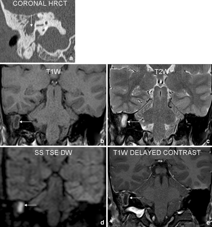
Fig. 1.
Case 1: 23 year old male patient with history of right sided conductive hearing loss and purulent discharge. Coronal HRCT (a) shows a soft tissue density lesion (arrow) involving right mastoid–middle ear complex with irregular bony destruction. Focal area of signal abnormality in right mastoid (arrows) on T1W (b) and T2W (c) coronal images shows diffusion restriction (appears bright) on SS TSE DW (d) sequence. Delayed postcontrast (e) images reveal non-enhancing nature of the lesion with mild peripheral rim enhancement. The findings were interpreted as positive for cholesteatoma. Surgery revealed right sided cholesteatoma

