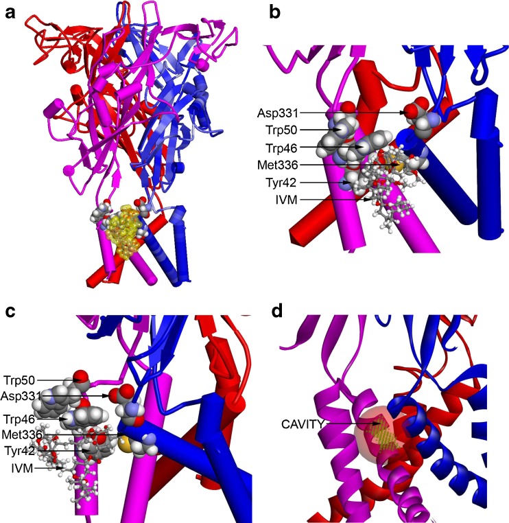
Fig. 6.
Homology model of rat P2X4R. (a) A homology model of rat P2X4R was built by threading the rat primary sequence onto the recent X-ray structure of the zebrafish P2X4R in the open conformation (PDB ID 4DW1) as described in “Materials and methods”. In this model, the P2X4R subunits are rendered as blue, pink, and red cartoon structures. The residues important for IVM binding are rendered with a van der Waals surface (carbon, oxygen, nitrogen, and hydrogen are colored gray, red, blue, and white), whereas the IVM is rendered in ball and stick and then enveloped with a transparent van der Walls surface (yellow). (b) A detailed view of the region surrounding the IVM molecule in Fig. 6a showing the relationship of residues important for IVM and ethanol binding. As viewed from the plane of the membrane and looking toward the ion pore, the IVM is between two subunits and within the membrane, but near the interface between the extracellular and transmembrane domains. The initial position of the IVM molecule was parallel to the plane of the membrane, as it is in the X-ray structure of GluCl. (c) A view of the region surrounding the IVM molecule in Fig. 6b, but rotated 90° in order to show the relationship of residues important for IVM and ethanol binding. The view is from the plane of the membrane but tangential to the ion pore, which is aligned with the right edge of the figure. The IVM molecule is between Trp46 and Met336; it also forms an H bond with Tyr42. (d) The IVM molecule was removed from the model in Fig. 6a and a cavity-finding algorithm was used to detect internal and external cavities. The module found 39 cavities using a 0.5-Å3 grid. The most relevant cavity site (to be compared to the IVM surface in Fig. 6a) is shown as a green grid enclosed within a transparent red sphere. It is located between two P2X4R subunits and is near the extracellular domain. In this figure, the P2X4R subunits are rendered as blue, pink, and red ribbons to better reveal the cavity

