Fig. 3.
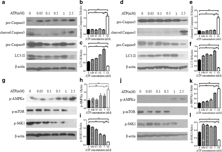
Effect and signaling transduction of eATP on anchorage-independent hepatoma cells. a–f BEL7402 hepatoma cells were seeded into poly-HEMA-coated six-well plates as anchorage-independent hepatoma model, and increasing concentrations of eATP (0–2.5 mM) were added to cells simultaneously (a–c) or after incubation for 24 h (d–f). Cells were incubated for another 12 h, and activation of caspase 3, caspase 9, and LC3-II was analyzed by Western blot (a and d). Caspase 3 (b and e) and LC3-II (c and f) bands were quantified densitometrically using Image J software. g–l In these two anchorage-independent models, activation of mTOR and AMPK signaling pathways was analyzed by Western blot, and β-actin was used as a loading control (g and j). Relative levels of p-AMPKα (h and k) and p-S6K1 (i and j) were quantified by densitometric analysis and normalized to β-actin. Presented figures are representative data from three independent experiments. *P < 0.05; **P < 0.01. NS not statistically significant

