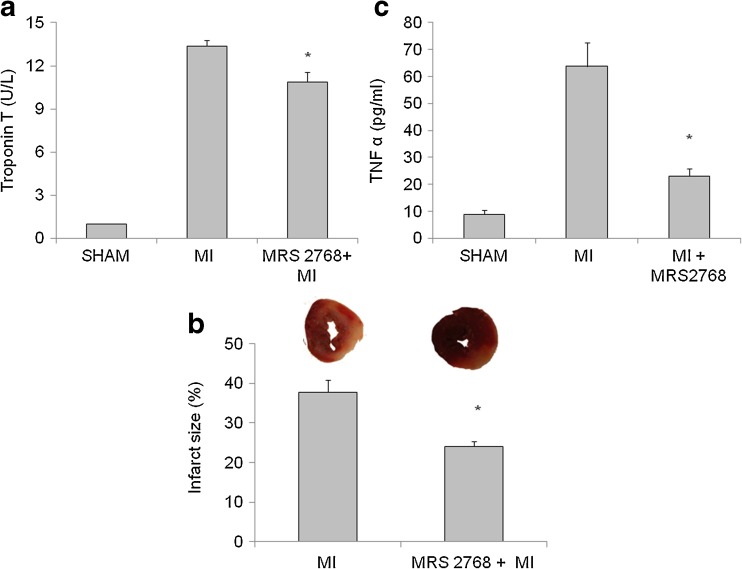
Fig. 4.
Measurements of Troponin T, infarct size, and TNF-α. a The release of Troponin T to the serum 24 h post-LAD ligation. Values are means ± SE. n = 12 hearts in each group. *p < 0.05 vs. MI. b The percent of irreversible injury was determined by scanning the images of mice hearts ventricular sections with triphenyl tetrazolium chloride (TTC). Representative images of the two different groups, revealing different degrees of myocardial ischemia (white to red zones after TTC staining). Hearts were subjected to 24 h of LAD ligation. A significant size of damaged tissue was noted in the myocardium of MI group. Mice pretreated with MRS2768 had significantly smaller infarct size. This figure represents the percent of irreversible injury from the total area of the section at 24 h post-LAD ligation. Values represent means ± SE. n = 7 hearts in each group. c The levels of TNF-α in the serum 24 h post-LAD ligation. Values are means ± SE. n = 4 in each group. *p < 0.05 vs MI

