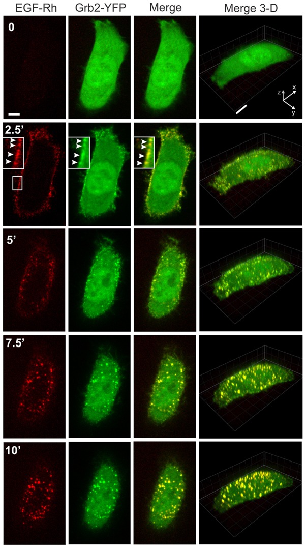
Fig. 1.

Time-lapse imaging of Grb2–YFP in cells stimulated with 2 ng/ml EGF–Rh (0–10 minutes). HeLa-Grb2–YFP cells were examined by 3D time-lapse imaging during the first 10 minutes after injection of 2 ng/ml EGF–Rh into the stage chamber at 37°C with 30-second intervals between frames as described in the Materials and Methods. Selected x–y time frames are presented (see corresponding supplementary material Movie 1). Each x–y image of EGF–Rh (red), Grb2–YFP (green) and the merged fluorescence (Merge) is the maximum projection of three neighboring confocal sections. Corresponding merged Rhodamine and YFP images are presented as the complete 3D x–y–z images. Insets are high magnification images of the region indicated by white rectangles. White arrowheads point on the examples of colocalization of EGF–Rh and Grb2–YFP in the plasma membrane. Scale bars: 5 µm.
